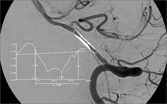
Digital Subtraction Angiography (DSA)
- Qualitative diagnosis of cerebrovascular diseases
- Location of lesion Lumen diameter of the narrowest lumen and reference vessels before endovascular treatment
- Lumen diameter of the narrowest and reference vessels after endovascular treatment Length of stenosis
- Detail description of lesions such as calcification analysis, intraplaque hemorrhage, and plaque rupture
- Length of stenosis
- Description of cerebrovascular anatomic variation
Magnetic resonance image (MRI)
- Accurate localization of cerebral infarction
- Mechanism analysis of cerebral infarction
- The number and volume of cerebral infarction
- Analysis of blood supply vessel for the district of cerebral infarction

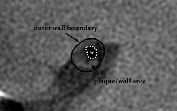
High-Resolution Magnetic Resonance Imaging (HRMRI)
- Lesion location on the coronal position such as internal carotid artery (ICA), middle cerebral artery (MCA), and basilar artery (BA)
- Lesion location on the axial position such as ventral, dorsal, and side location of the plaque
- The condition of the plaque surface
- Single or tandem lesions
- Length of plaque, thrombus, or occlusion
- Plaque burden
- Remodeling index
- Hyperintense plaque (HIP)
- Enhancement index
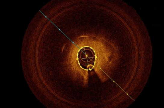
Optical Coherence Tomography (OCT)
- Interpretation of patch crack (fiber cap continuity)
- Determination of fiber cap thickness
- Determination of the degree of lumen stenosis
- Interpretation of calcification degree and pattern (nodular vs. dispersed, superficial vs. deep), and determination of calcification index
- Interpretation of intraluminal thrombus or plaque erosion
- Interpretation of neovascularization or vasa vasorum
- Detailed description of target vessels after endovascular treatment such as the stent position

Computational Fluid Dynamics (CFD)
- The diameter of the narrowest and reference vascular vessels, and the decrement proportion of distal diameter
- Wall Shear Stress (WSS) and Unit Time Wall Shear Stress
- Oscillatory Shear Index (OSI) and Relative Retention Time (RRT)
- Fractional Flow Reserve (FFR)
- Reduction coefficient of blood pressure and downstream vascular impedance
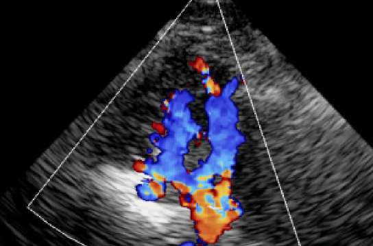
Vessel Ultrasound (VU)
- Interpretation of plaque properties
- Medial thickness at the narrowest site
- Lumen diameter of the narrowest and reference vessels
- Lumen area of the narrowest and reference vessels
- Flow velocity at systolic peak and diastolic peak
- Resistance index
- Variation of the frequency signal